life in the fast lane ecg pdf
Lead PlacementLead Placement V1 4th ICS i ht tth ICS right sternum V2 4th ICS left sternum V3 midway between V midway between V2. AV-nodal reentry tachycardia SVT 3.

Killer Ecg Patterns Part 1 Litfl Ecg Library
Hard to interpret an ECG with LBBB Lead V1 Q wave and an S wave Lead V6 an R wave followed by another R wave Lead V6 Rabbit ears.

. The earliest manifestation of hyperkalaemia is an increase in T wave amplitude. Some ECGs have been obtained from Derriford Hospital Plymouth. 2nd degree Mobitz I Dec 11 2021 LITFL-Life-in-the-FastLane-760-180 The first clue to the presence of Mobitz I AV block on this ECG is the way the QRS complexes cluster into groups separated by short pausesThis phenomenon usually represents 2nd-degree AV block or MBBS CCPU RCE Biliary DVT E-FAST AAA Rob is an.
Sep 25 2021. I used this resource later in my studies when i already had some basic knowledge. .
1 Progressive PR interval. Supraventricular Tachycardia Life in the Fast Lane ECG Library Narrow complex tachycardia at 125 bpm. ECG Axis Interpretation From Life In The Fast Lane Posted on May 31 2019 by Tom Wade MD I will be erasing the post below and changing it to the title blog post.
Images from other websites are free to use under Creative Commons Attribution Non-Commercial Share Alike 30 and 40 licenses. Abdominal Pain afibrvr Allergic Reaction Anaphylaxis Spectrum Altered Mental Status - Intoxication AMA. 2 Progressive RR interval.
Name Email Website. Inverted in II III and aVF. Remember early repol is called early repol because repolarization comes early relatively short QT Third there is.
Life threatening arrhythmias. Atrial flutter with 21 block especially in elderly IHD CCF 2. Litfl is a medical blog and website dedicated to providing online emergency medicine and critical care insights and education for everyone everywhereusually Ecg axis interpretation from life in the fast.
What is the diagnosis of this ECG. Mobitz I ECG Characteristics Life in the Fast Lane ECG Examples. Life in fast lane ecg axis.
S1Q3T3 pattern in ECG is seen in acute pulmonary embolism 1. ECG Reference SITES and BOOKS the best of the rest. EKGECG rhythm template from Life in the Fast Lane EKGECG rhythm template from Life in the Fast Lane ekg rhythm templatepdf.
Life in the fast lane ecg videos. What are the differentials for a narrow complex tachycardia. Second the QT appears slightly long for early repol.
It is very rare to have non-concavity convex or straight in any one of leads V2-V6 in normal variant ST elevation. All images have been referenced - to see the reference click on the image. 2 Q H 1 R P O L K P S V R Q H I L F M O Y U 6 A X B 0.
Hyperkalaemia is defined as a serum potassium level of 52 mmolL. S1Q3T3 pattern is the classical ECG pattern of acute pulmonary embolism which is often taught in ECG classes though it is not the commonest. 4 RR interval containing the non-conducted P wave than the sum of RR interval prior to the pause b.
Retrograde P waves follow each QRS complex. Life in the fast lane. ECG A to Z by diagnosis ECG interpretation in clinical context.
Identify ECG changes related to hypertrophy bundle branch blocks and MIs Review approach to interpretation of wide complex tachycardia Describe other miscellaneous causes of ECG abnormalities. S1Q3T3 pattern means the presence of an S wave in lead I indicating a rightward shift of QRS axis with Q wave and T inversion in lead III. Life in fast lane ecg lbbb.
Pericarditis electrolyte abnormality medication effects and hypothermia Practice using a systematic approach to interpreting 12 lead. ECG Exigency and Cardiovascular Curveball ECG Clinical Cases. EraC lacitirC dna etucA ycnegremE ni yhpargoidracortcelE 7102 IM etucA ni GCE ehT eraC lacitirC dna enicideM ycnegremE ot tnaveler scipot GCE 001 revo gnirevoc ecruoser lanoitacude eerf a si yrarbil GCE LFTIL gnidaeR rehtruF LFTIL FDP 2002 ikiW enicideM ycnegremE labolG ehT.
100 ECG Quiz Self-assessment tool for examination practice. When you see a regular narrow complex tachycardia at 150 bpm you should think of four main diagnoses. First as you said there is a nearly straight ST segment.
2nd Degree Heart Block If only there is only 21AV blocks on the ECG one cannot distinguish between Mobitz I from Mobitz II. ECG changes generally do not manifest until there is a moderate degree of hyperkalaemia 60 mmolL. Lane Lane 1 Lane 23 Lane 45 Lane 6 Lane 7 Distribution Weibull Log-Logistic Weibull Exponential Log-Logistic R 2 0667377 0421123 03203 0542482 0667383.
Leave a Comment Cancel reply. ECG Library Basics Waves Intervals Segments and Clinical Interpretation.

Ekg Basics Litfl Litfl Ecg Library
Ekg Basics Litfl Litfl Ecg Library

100 Ecg Quiz Self Assessment And Clinical Cases Litfl Education Blog Self Assessment Emergency Medicine

Learn Ecg In A Day Pdf Am Medicine Nursing Books Icu Nursing Nursing School Survival
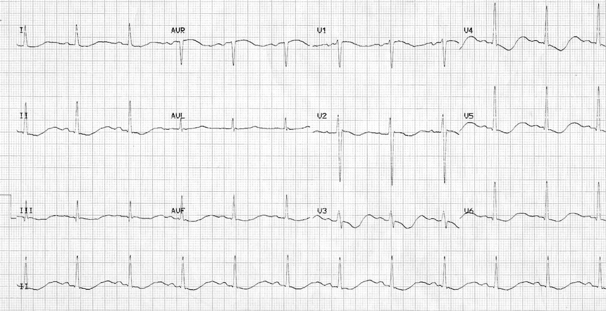
Hypokalaemia Ecg Changes Litfl Ecg Library
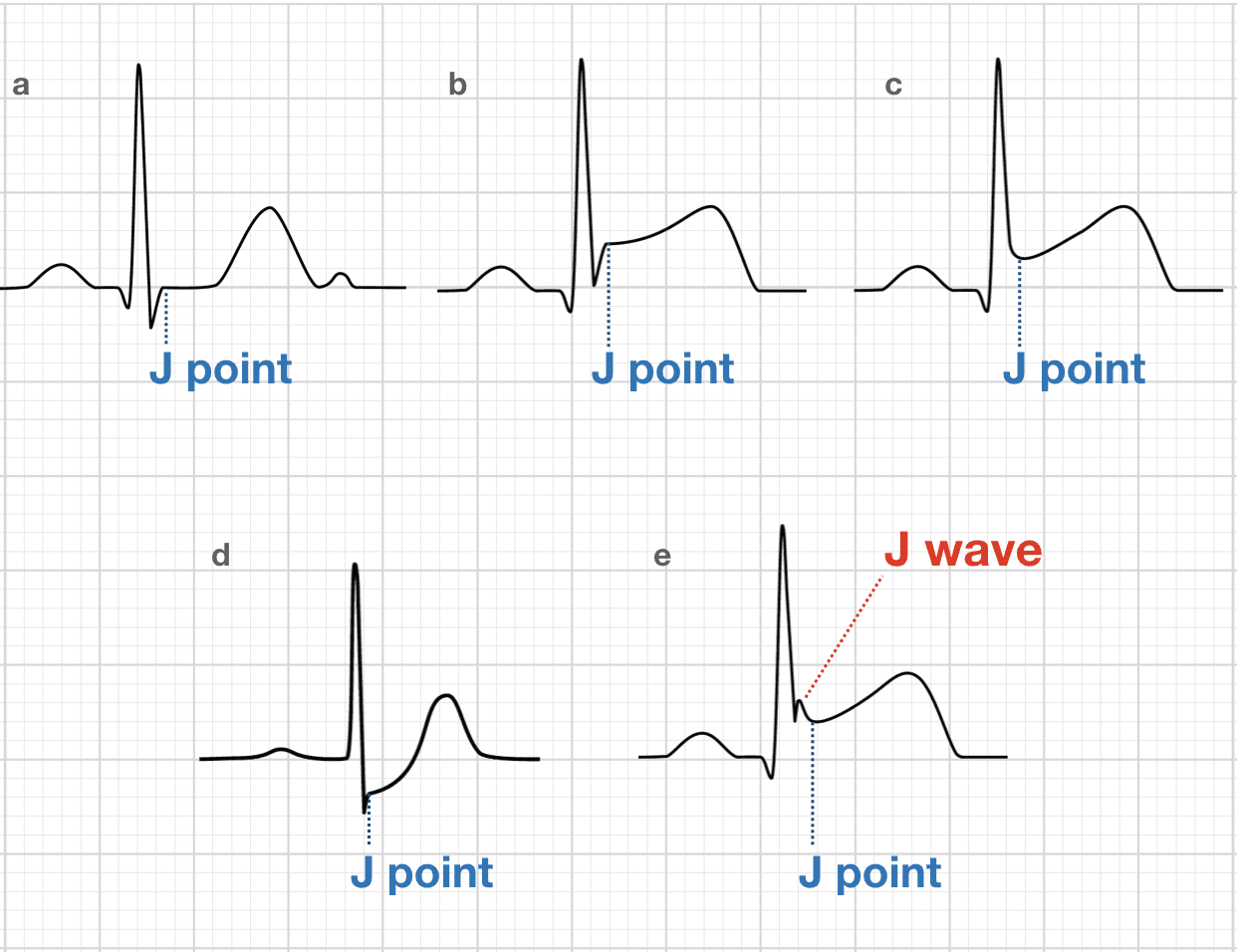
J Point Ecg Interval Litfl Ecg Library Basics

Ecg Interpretation Cheat Sheet Download As Pdf File Pdf Text File Txt Or Read Online Ecg Interpretation Cheat Sheets Cheating

12 Lead Ecg Interpretation Lesson And Practice Quiz 311 Ekg Interpretation Ecg Interpretation Emergency Nursing
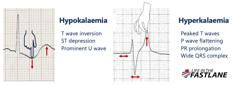
Hyperkalaemia Ecg Changes Litfl Ecg Library

Ecg Left Ventricular Hypertrophy Lvh Emt Paramedic Segmentation Nursing Tips

Ecg Rate Interpretation Litfl Medical Blog Ecg Library Basics

Right Axis Deviation Ecg Rad Litfl Ecg Library Ecg Interpretation Cardiology Nursing Nursing Study
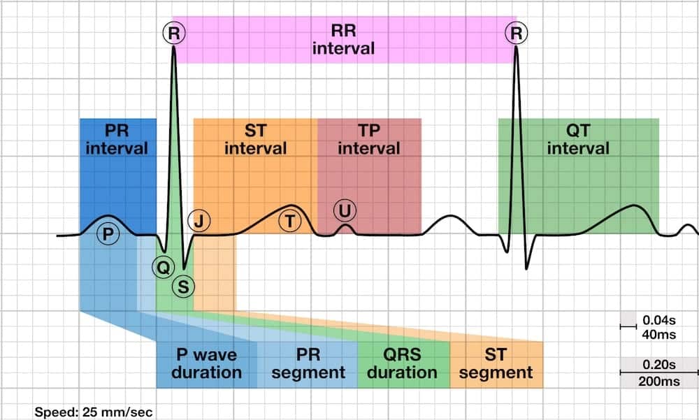
Pr Interval Litfl Ecg Library Basics
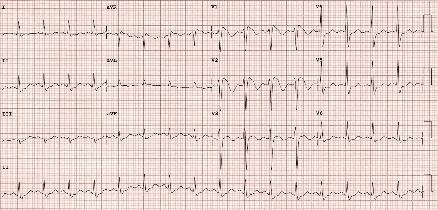
Brugada Syndrome Litfl Ecg Library Diagnosis

Pin On Education Books That Get You Thinking

Ecg Left Ventricular Hypertrophy Lvh Emt Paramedic Segmentation Nursing Tips

Right Axis Deviation Ecg Rad Litfl Ecg Library Ecg Interpretation Cardiology Nursing Nursing Study

